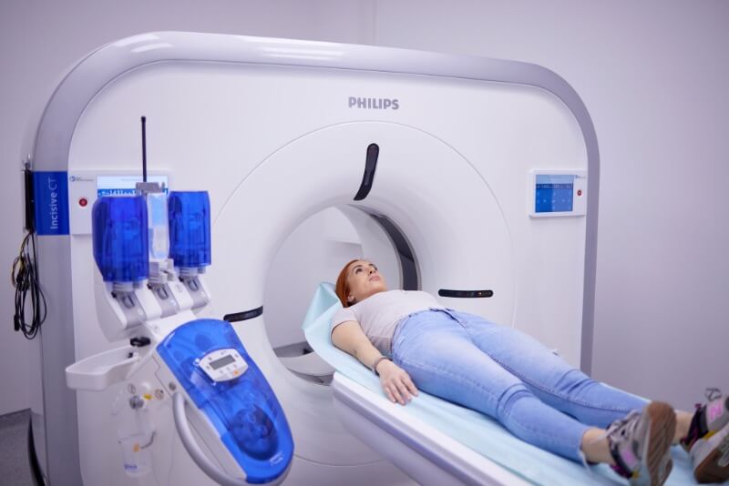
What is a CT Scanner?
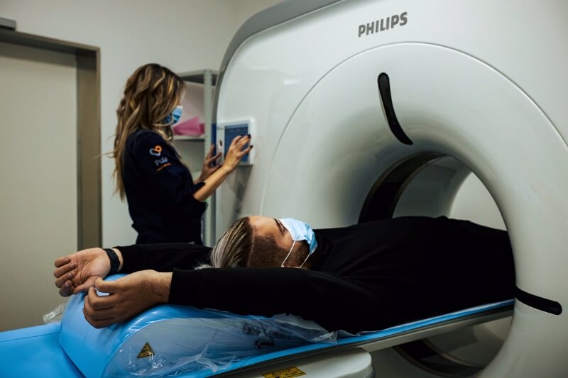
A CT scanner (CT, computed tomography) is a medical imaging device that uses X-rays and computer technology to create detailed images of the inside of the human body. CT scanners are used to diagnose various diseases and conditions because they can detect and display small changes in the body that are not visible on regular X-ray images.
The CT scanner works by rotating an X-ray tube around the patient, emitting directed X-rays through the body. Detectors on the opposite side of the body then measure how much of the X-rays have passed through the tissue. A computer processes this data to create 2D or 3D images of the body’s interior. The resulting images show detailed cross-sections of the body, including organs, bones, and soft tissues, providing valuable diagnostic information to healthcare professionals.
Which Conditions Can Be Diagnosed with a CT Scan?
CT scanners play a crucial role in diagnosing various medical conditions and diseases because they can produce detailed images of the body’s internal structures. CT scans are used to diagnose:
Assessment of Heart and Blood Vessel Health
A heart CT scan provides a detailed view of the heart’s structures. A CT Calcium Score is used to assess the presence and extent of calcifications in the coronary arteries.
Evaluation of Coronary Artery Disease
CT coronary angiography is used for diagnosing, monitoring, and planning surgical treatment in patients with coronary artery disease, as well as for assessing the risk of heart attack. For a more precise evaluation of hemodynamic impairment in coronary arteries, MDCT FFR (Fractional Flow Reserve) is used.
Assessment of the Aorta
A CT scan of the aorta is used to evaluate aneurysms, dissections, and congenital anomalies of the aorta.
Detailed Examination of Blood Vessels
CT angiography is used to examine the blood vessels of the brain, neck, chest, abdomen, pelvis, legs, and arms.
For Acute Chest Pain
TRO CT angiography (Triple Rule-Out) helps exclude life-threatening causes.
Diagnosing and Monitoring Lung Diseases
A lung CT scan can detect lung conditions such as pneumonia, emphysema, and lung cancer.
Diagnosing Injuries
CT scans are commonly used to diagnose and assess injuries and fractures. Depending on the location of the injury, the following scans may be performed:
-
CT scan of the head
-
CT scan of the chest
-
CT scan of the abdomen and pelvis
-
CT scan of the spine
-
CT scan of the joints
This includes evaluating the severity of bone fractures, detecting internal bleeding, and assessing organ damage.
Diagnosing Degenerative Diseases
-
Head CT for Alzheimer’s disease
-
Spinal CT for herniated discs
-
Spinal stenosis
-
Joint CT for osteoarthritis
Assessment of Cerebrovascular Diseases
A head CT is used to diagnose and monitor strokes or intracranial bleeding.
Diagnosing and Monitoring Inflammatory Conditions
-
Head CT for evaluating meningitis and encephalitis
-
Joint CT for rheumatoid arthritis
-
Spinal CT for osteomyelitis
Assessment of Abdominal Organs
CT of the abdomen and pelvis provides detailed imaging of the liver, pancreas, kidneys, spleen, and intestines.
Assessment of the Urinary Tract
CT urography is used to diagnose kidney stones, inflammatory conditions, abnormalities, and tumors of the urinary tract.
Detecting Cancer
CT scans are frequently used to detect tumors and cancerous growths in various parts of the body, including the lungs, liver, and pancreas.
Before Scheduled Surgery
CT scans are performed before surgery to determine the extent of the procedure and help plan postoperative care.
CT scanners play a crucial role in diagnosing a wide range of medical conditions and diseases, enabling healthcare professionals to make informed decisions about the best course of treatment for their patients.
What Is a CT Scan with Contrast?
A CT scan with contrast is a diagnostic imaging technique that uses X-rays along with a contrast agent to produce detailed images of the body’s interior. The contrast material, which usually contains iodine, is injected into a vein in the arm or leg. Oral and rectal contrast agents may also be used, depending on the procedure.
What Is Contrast?
Contrast enhances the quality of CT images by making it easier to distinguish different tissues and structures inside the body. The contrast agent contains a chemical element that absorbs X-rays more effectively than surrounding tissues, resulting in images with greater clarity. As the contrast travels through the bloodstream, it accumulates in specific tissues or organs, making them more visible on the resulting images. The use of contrast can help detect medical conditions that may not be visible on a non-contrast CT scan.
CT scans that may use contrast include:
-
CT angiography – used to visualize blood vessels throughout the body
-
CT urography – used to visualize the urinary tract
-
CT pulmonary angiography – used to visualize the pulmonary arteries
These are just a few examples of the many types of contrast-enhanced CT scans available, each tailored to the patient’s specific needs and condition.
How Should I Prepare for a CT Scan?
Preparation for a CT scan may vary depending on the body part being scanned and the type of scan being performed. Here are the general steps to follow before a CT scan:
-
Inform your doctor of any allergies, medical conditions, or medications you’re taking—especially those that may affect your kidneys or thyroid function.
-
Follow all instructions from your doctor regarding fasting, medications, or other preparations. For example, you may be asked not to eat or drink for a certain period before the scan or to stop taking certain medications that could affect the results.
-
Wear comfortable clothing: Wear loose, comfortable clothes without any metal parts such as belts or jewelry.
-
Remove metal objects: Take off any metal items such as dentures, hearing aids, or piercings, as they can interfere with the scan.
Are There Any Restrictions or Requirements for a CT Scan?
Contrast agents may be contraindicated for patients with kidney disease or allergies to iodine or contrast materials. Certain CT scans—especially of the abdomen or pelvis—may require fasting for several hours beforehand to ensure clear imaging of the area being scanned. Some medications, such as anticoagulants or insulin, may need to be adjusted or paused before the scan to reduce the risk of complications.
Some patients may experience claustrophobia or anxiety during the scan, particularly when it involves entering a narrow or enclosed space. It’s important to note that pediatric patients are more sensitive to radiation exposure, and CT scans are generally not recommended for pregnant women, especially during the first trimester.
However, in some cases, the benefits of the scan may outweigh the risks, and the healthcare provider will make that decision on a case-by-case basis. Some patients may require special preparation or alternative diagnostic tests.
By following these general guidelines, you can help ensure that your CT scan is safe, accurate, and effective in diagnosing any potential health condition.
How Long Does a CT Scan Take?
The duration of a CT scan depends on the type of scan being performed. A standard CT scan without contrast typically takes around 10–15 minutes. However, some procedures—such as CT angiography of the heart—can take up to 30–40 minutes.
If a contrast agent is required during the scan, the process may take longer—usually 30–60 minutes—as the contrast material is administered intravenously before the scan begins.
The total time can also vary depending on several factors, including:
-
The area being scanned
-
The complexity of the procedure
-
The patient’s condition and cooperation
-
Technical issues that may arise during the scan
These factors can influence the overall duration of the examination.
Overview of a Typical CT Scan Process:
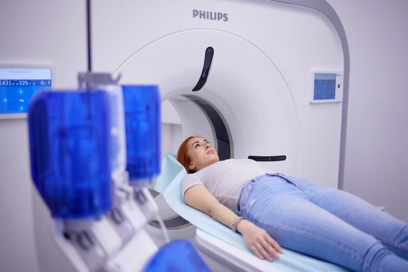
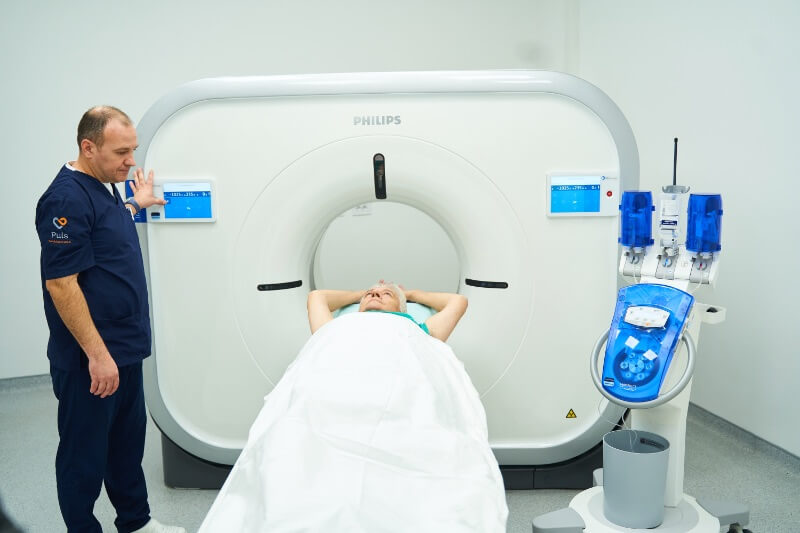
- Positioning: You will lie on a flat table that slides into the CT scanner.
- Scanning: The CT scanner will rotate around your body, capturing multiple X-ray images from different angles. You may hear buzzing or clicking sounds during the scan, but you won’t feel any pain.
- Contrast Injection (if needed): A contrast agent may be injected into a vein in your arm or hand during the scan.
- Completion: Once the scan is finished, you’ll be moved out of the machine, and the healthcare provider will remove any IV lines or positioning aids.
- Monitoring: A medical professional will review the images and provide a diagnosis or recommend further testing or treatment if necessary.
It is important to follow all instructions or preparations provided by your healthcare provider to ensure the scan is safe and effective, and to report any concerns or discomfort you may feel during the procedure.
Some of the CT Scans Performed at the Puls Dedinje Emergency Center Include:
- CT scan of the head
- CT scan of the spine
- CT scan of the chest
- CT scan of the hips
- CT scan of the abdomen
- CT scan of the pelvis
- CT scan of soft tissues
- CT scan of the thigh
- CT scan of the shoulder
- CT scan of the upper arm
- CT scan of the foot
- CT scan of the orbits (eye sockets)
- CT scan of joints
- CT scan of the lower leg
- CT scan of the sinuses
What Does a CT Scan Show?
A CT scan produces a series of X-ray images processed by a computer to create detailed cross-sectional images of the inside of the body. These images are usually displayed on a computer monitor or printed on film for further analysis by a medical professional. CT scans help diagnose a wide range of medical conditions and can reveal:
- Internal bone structures
- Tumors
- Soft tissue injuries
- Blood vessel problems
- Infections
- Trauma
- Organ issues
- Other abnormalities
Types of images a CT scanner can provide include:
- Axial images: Show detailed cross-sections of internal structures such as organs, bones, or soft tissues.
- Sagittal images: Show the body’s internal structures in a vertical plane.
- Coronal images: Display the body’s internal structures in a horizontal plane.
- Three-dimensional images: Combine multiple image slices to create a 3D view of internal structures, useful for surgical planning or visualization.
- Functional images: Provide information about the function or activity of certain internal structures, such as blood flow, perfusion, or metabolism.
CT (Computed Tomography) Scans Can Visualize a Wide Range of Body Structures and Conditions:
- Heart, lungs, and blood vessels: Used to diagnose and monitor coronary artery disease, heart attacks, and vascular blockages.
- Bones, joints, and spine: Used to diagnose fractures, arthritis, bone tumors, joint diseases, herniated discs, or spinal stenosis.
- Soft tissues: Such as muscles, organs, and blood vessels, for diagnosing cancer, infections, or inflammation.
- Brain: Helps in diagnosing and monitoring strokes, brain hemorrhages, or brain tumors.
- Lungs: To diagnose conditions like pulmonary embolism, pneumonia, or lung cancer.
- Abdominal and pelvic organs: Including the liver, spleen, pancreas, kidneys, uterus, ovaries, and prostate — used to diagnose tumors, infections, or cysts.
- Blood vessels throughout the body: To diagnose conditions such as aneurysms, blood clots, or arterial blockages.
Can I Undergo a CT Scan If I’m Pregnant?
CT scanning is generally not recommended during pregnancy, especially in the first trimester. The use of a CT scanner during pregnancy may expose the fetus to X-rays, which are considered potentially harmful to fetal development. Radiation can damage cells and cause genetic mutations in the fetus, increasing the risk of abnormalities such as cancer or other health issues.
However, in certain urgent cases, when imaging is essential, a doctor may determine that the potential benefits outweigh the risks and recommend a CT scan.
If a CT scan is necessary, several precautionary measures can be taken to reduce the risk to the fetus, such as:
-
Using the lowest possible radiation dose
-
Providing protective equipment for pregnant women, such as lead aprons
Always consult with your doctor to evaluate the necessity and safety of a CT scan during pregnancy.
What Is an Alternative Option for Pregnant Women?
One of the most common alternatives to a CT scan for creating detailed images of internal organs and body structures is ultrasound, which uses sound waves, and magnetic resonance imaging (MRI), which uses a magnetic field and radio waves. Both methods are painless, non-invasive, and do not involve the use of X-rays, making them safe for pregnant women.
It is very important to talk to your healthcare provider about any concerns you have regarding CT scans during pregnancy. Your doctor will assess your health, pregnancy status, and inform you about the risks and benefits of a CT scan, as well as available alternative diagnostic methods that are safe during pregnancy. If your doctor determines that a CT scan is necessary, they will provide you with information on how to protect yourself and minimize any risks to your baby.
Is a CT Scan Dangerous?
CT scans are considered safe and effective diagnostic tools, but like any medical test, it’s important to carefully weigh the risks and benefits and consult your doctor. Following all instructions given before, during, and after the scan helps ensure the best possible results and minimizes potential risks.
CT scanning is generally regarded as a safe diagnostic procedure, but there are potential risks and side effects to consider:
-
Radiation exposure: A CT scan uses X-rays, which can slightly increase the risk of cancerous cell changes. However, the radiation dose used is typically low, and healthcare providers take care to minimize the patient’s exposure as much as possible.
-
Allergic reactions to contrast agents: It’s important to inform your doctor if you have a history of allergies or sensitivity to medications or contrast materials.
-
Claustrophobia (fear of enclosed spaces): Some patients may feel discomfort during the scan due to the enclosed environment.
Ways to Ensure CT Scan Safety Include:
Using the minimum radiation dose
Medical professionals aim to reduce the amount of radiation used to the lowest effective level.
Using protective equipment
When contrast material is used, patients are usually provided with protective gear, such as a lead apron, to help reduce radiation exposure.
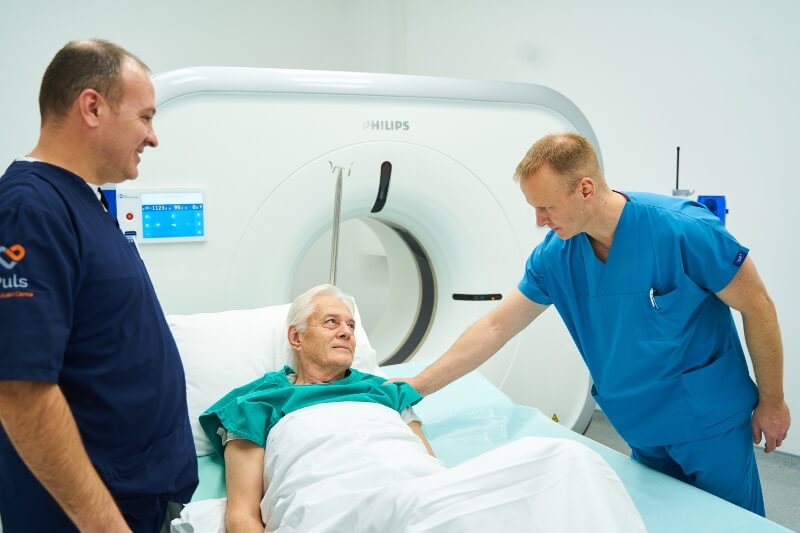
Patient Monitoring
Healthcare professionals monitor patients during a CT scan to ensure that no side effects or adverse reactions occur. If any unwanted reaction is observed, the doctor will act quickly to prevent further complications.
Use of Contrast Material
Contrast material can enhance image quality, but it is used only when necessary. Doctors will evaluate the benefits and risks of using contrast before making a decision, especially if the patient has a history of allergies to medications or contrast agents.
What Are Common Conditions That Can Be Detected with a CT Scan?
A CT scan can detect a variety of conditions and pathologies in the body, including:
- Cancer in different parts of the body
- Blood clots
- Bone and joint damage
- Heart and internal organ problems
- Infections
- Inflammations
- Various other abnormalities
The CT Scanner is a very useful diagnostic tool that can help detect and monitor conditions that have already been diagnosed. For example, when a tumor is discovered, a CT scan can be used to determine its size and location, as well as to monitor its growth or changes over time. A CT scanner can also be used to track how a tumor responds to treatment, helping to evaluate the success of the therapy.
Additionally, a CT scan can assist in detecting and monitoring other conditions, such as inflammation and infections. For instance, if pneumonia is suspected, a CT scan can help confirm the diagnosis and monitor the progress of treatment and recovery. In cases of bone injuries, a CT scan can help detect fractures and other bone issues, as well as monitor the healing process over time.
CT scanning aids in diagnosing and monitoring various conditions and pathologies in the body, allowing doctors to provide more accurate and effective diagnoses and treatment plans.
When Should You Contact Your Doctor After a CT Scan?
Signs and symptoms that may require medical attention after a CT scan include:
- Allergic reactions to contrast material, such as rash, itching, swelling, or difficulty breathing
- Nausea and vomiting
- Headache
- Pain or swelling at the injection site
- Concerns about scan results or the procedure itself
If you notice any of these symptoms after a CT scan, inform your healthcare provider. In some cases, these symptoms can be serious and require medical attention. It’s important to follow your provider’s instructions on how to take care of yourself after the scan and to contact your doctor if you experience any unusual symptoms or side effects.